Orthopedic surgery solves a mechanical problem; rehabilitation is what turns that mechanical solution into a functional recovery. The difference between a dog that returns to full activity in 16 weeks and one that plateaus at 70 percent function rests almost entirely on what happens between the surgeon's work and the owner's first post-op walk around the neighborhood. This article lays out a week-by-week rehabilitation framework consistent with published protocols from multiple veterinary teaching hospitals, including the University of Tennessee, Colorado State University, and the Canine Rehabilitation Institute.
Why Timelines Matter
Biological healing follows predictable phases: inflammation, proliferation, and remodeling. The inflammatory phase dominates the first 3 to 7 days, the proliferative phase covers roughly week 1 through week 4, and the remodeling phase extends from week 4 through six months or more depending on the tissue involved. Rehabilitation interventions that are appropriate in one phase can be harmful in another. Aggressive resistance exercise during the inflammatory phase, for instance, delays healing and increases the risk of re-injury. Conversely, prolonged rest into the remodeling phase produces muscle atrophy and joint stiffness that can become permanent.
The American Animal Hospital Association's rehabilitation guidelines emphasize that time-based progression must be combined with criteria-based progression. The calendar tells us what interventions are probably safe; the dog's clinical response tells us whether those interventions should actually be applied this week.
Weeks 1 to 2: Protected Inflammation
During the first two weeks, the surgical site is in active inflammation, and fixation hardware is bearing most of the mechanical load. The rehabilitation priority is pain management, swelling control, and preservation of joint range of motion in joints adjacent to the surgical site. Cryotherapy is applied 3 to 4 times daily for 10 to 15 minutes. Passive range-of-motion exercises are performed gently through pain-free range, typically two sessions per day of 10 to 15 repetitions per joint. Weight-bearing is limited to short, controlled leash walks of 3 to 5 minutes, typically two or three times a day, on soft non-slip surfaces.
Our detailed breakdown of gentle joint mobilization is covered in our range of motion exercises guide. For dogs that underwent TPLO specifically, the week-by-week adjustments are documented in our TPLO surgery recovery timeline.
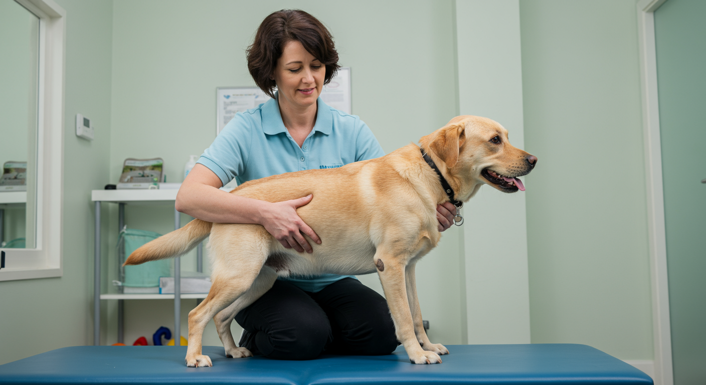
Weeks 3 to 4: Early Active Rehabilitation
Suture removal typically occurs around day 10 to 14, marking the transition from protected inflammation to early active rehabilitation. Underwater treadmill therapy becomes appropriate once the incision is watertight, which most surgeons clear between days 14 and 21. Proprioceptive exercises can be introduced, starting with slow weight shifts on a stable surface. Sit-to-stand repetitions and three-leg standing exercises begin to rebuild muscle activation patterns around the surgical site.
Exercise duration during this phase usually progresses from 8 minutes to 15 minutes of active rehabilitation per session, with two sessions per day. Leash walking extends to 10-minute blocks performed on flat ground, still on a short leash to prevent sudden movements. Any increase in limb use or lameness at the end of a session is a signal to scale back before progressing further.
Weeks 5 to 8: Progressive Strengthening
This phase focuses on rebuilding strength, endurance, and coordination. Dogs that have shown steady progress through the early active rehabilitation phase begin functional exercises such as controlled stair work (3 to 5 steps, assisted), gentle hill walking, and cavaletti (ground poles) to challenge limb awareness. The balance and proprioception training protocols become central to this phase. Underwater treadmill sessions increase to 15 to 20 minutes, with gradual reduction in water depth to increase weight-bearing load.
Published outcome data supports the value of this phase. A 2015 study in Veterinary Surgery by Monk and colleagues found that dogs receiving structured rehabilitation after TPLO regained thigh circumference symmetry significantly faster than dogs managed with activity restriction alone. The practical implication is that owners willing to invest in this phase usually see their dogs walking more symmetrically, with fewer compensatory patterns, by the end of week 8.
Case Example: Dexter, TPLO Recovery
Dexter, a 4-year-old Labrador, underwent right-side TPLO after a cruciate ligament rupture. His rehabilitation followed the framework outlined here. Thigh circumference measured weekly showed 2.3 cm deficit at week 2, 1.6 cm at week 6, and full symmetry by week 10. Force plate analysis at week 12 showed 96 percent symmetry in peak vertical force, compared with an average of 82 percent in a matched cohort of dogs managed without rehabilitation. His return to full off-leash activity occurred at week 14, consistent with the published return-to-sport literature on structured post-op protocols.
Weeks 9 to 16: Functional Return
The final phase bridges clinic-based rehabilitation and return to normal activity. Exercise variety increases: directional changes, controlled fetch with a short-rolling ball, swimming in open water if available, and gradually longer off-leash walks on safe terrain. Underwater treadmill sessions may taper to once weekly or every other week, with the focus shifting to land-based exercises that mimic normal activity patterns.
Return-to-sport dogs, including agility, field trial, and working service dogs, typically require an additional 4 to 8 weeks of sport-specific conditioning beyond the base rehabilitation program. Our progressive weight-bearing exercises article covers the late-phase strengthening work in more detail.
When Progress Stalls
Not every recovery follows the expected arc. Warning signs that warrant veterinary reassessment include persistent lameness beyond week 6, failure to regain thigh circumference within 10 percent of the contralateral limb by week 10, or sudden regression after previous improvement. Common reversible causes include over-exercise, inadequate pain control, or development of compensatory muscle tightness in the contralateral limb. Less common but more serious causes include implant failure, infection, or meniscal injury that was not apparent at the time of original surgery.
The American College of Veterinary Sports Medicine and Rehabilitation resources emphasize that experienced rehabilitation practitioners often detect subtle abnormalities before they become overt problems. Involving a Certified Canine Rehabilitation Practitioner or Certified Canine Rehabilitation Therapist, ideally from the day of suture removal onward, adds a layer of monitoring that catches these issues early.
Owner Contribution
Published compliance data suggests that owners complete 40 to 60 percent of prescribed home exercises on average, and this adherence strongly correlates with final outcomes. Simple tools improve compliance: a printed daily exercise log kept by the kitchen, a short video of each exercise filmed by the therapist at the clinic, and weekly phone or email check-ins with the rehabilitation team. Dogs whose owners are actively engaged in the home exercise program routinely outperform those whose owners rely solely on clinic visits.
A successful orthopedic rehabilitation is a partnership among surgeon, rehabilitation therapist, primary veterinarian, and owner, each contributing specific expertise at the right moments. When the partnership is functioning, most dogs reach within 5 percent of pre-injury function by week 16, and the majority return to the activities they enjoyed before surgery.