Canine neurological rehabilitation sits at the intersection of three disciplines: neurology, orthopedics, and motor learning. The central premise is that the damaged nervous system has more plasticity than traditionally believed, and that targeted, repetitive, task-specific movement promotes functional recovery even after substantial injury. A 2019 review in the Journal of Small Animal Practice by Sims and colleagues summarized mounting evidence that structured rehabilitation meaningfully improves ambulatory outcomes after intervertebral disc surgery, and clinical practice has followed suit. This article lays out the protocol frameworks used in contemporary veterinary rehabilitation for the three most common neurological patient populations.
Baseline Neurological Assessment
Rehabilitation cannot be planned without a clear picture of what the nervous system is still capable of. The standard assessment follows the Modified Frankel Scale or the Texas Spinal Cord Injury Score to grade motor function from non-ambulatory paraplegia through normal ambulation. Proprioceptive positioning, postural reactions, deep pain perception, and voluntary motor function are each evaluated and documented. The presence of deep pain perception is the single most important prognostic indicator for spinal cord injuries: dogs with intact deep pain have a 60 to 85 percent probability of regaining ambulation, whereas dogs with loss of deep pain for more than 48 hours carry a significantly guarded prognosis.
Muscle mass is measured by girth tape at standardized points along each limb, and gait is scored using the Olby Score (0 to 14), which captures ambulatory quality more precisely than binary ambulatory status. Reassessment at two-week intervals produces the objective trend data that owners rely on to judge whether the program is working.
Protocol 1: Post-Hemilaminectomy IVDD
Thoracolumbar intervertebral disc disease managed surgically with hemilaminectomy is one of the most common neurological indications for rehabilitation. The American College of Veterinary Internal Medicine consensus on IVDD managementrecommends early initiation of supportive care and, where facilities allow, rehabilitation beginning within the first week post-op.
During the first 2 weeks, sessions focus on passive range of motion for all four limbs (10 to 15 repetitions per joint, two to three times daily), manual bladder expression teaching for the owner, and assisted standing exercises using a sling. Electrical stimulation for muscle re-education may be introduced around day 7 to 10 depending on surgical clearance. Starting week 3, assisted walking with harness support, proprioceptive placement exercises, and underwater treadmill therapy become central. Water depth is kept high (shoulder level) initially to maximize buoyancy and allow voluntary stepping even in dogs with limited strength.
Progression from week 4 to week 12 typically shifts from passive to active exercises, from shorter to longer sessions, and from higher-buoyancy water to shallower water that gradually reloads the limbs. Our companion article on canine spinal injury rehabilitation covers the acute-phase considerations in more depth.
Protocol 2: Fibrocartilaginous Embolism (FCE)
FCE produces sudden, non-progressive spinal cord ischemia without surgical indication in most cases. Recovery is driven entirely by rehabilitation plus time. The protocol resembles the post-hemilaminectomy framework but begins immediately after diagnostic confirmation (typically MRI) rather than after a surgical recovery window. Dogs with preserved deep pain and asymmetric presentation (more common with FCE than with IVDD) often regain functional ambulation within 6 to 12 weeks with structured rehabilitation.
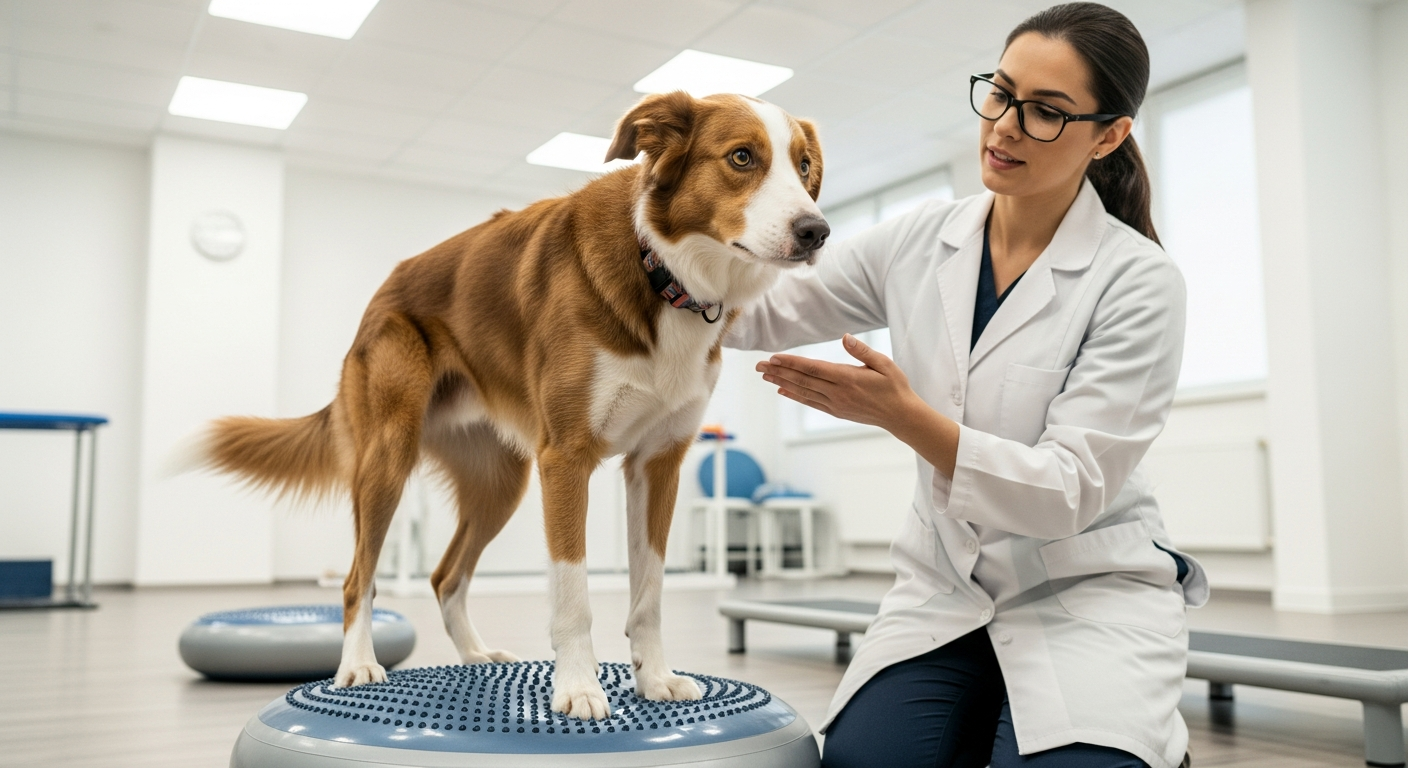
Key interventions include underwater treadmill therapy (2 to 3 sessions per week), balance work on foam pads and wobble boards, and cavaletti drills that force deliberate foot placement. Electrical stimulation for the most affected muscle groups during the first 3 to 4 weeks helps prevent atrophy while voluntary control is returning. Our balance and proprioception training article details the progression of these exercises.
Protocol 3: Degenerative Myelopathy
Degenerative myelopathy (DM), the canine analog of human amyotrophic lateral sclerosis, cannot be reversed, but a structured rehabilitation program has been shown to extend mean survival and preserve ambulation longer than supportive care alone. A 2006 study by Kathmann and colleagues found median survival of 255 days in DM dogs receiving physiotherapy versus 55 days in dogs without rehabilitation, a dramatic difference that has been reproduced in subsequent practice audits.
The DM rehabilitation protocol emphasizes maintenance of voluntary movement, weight control, and owner education about progressive assistive technology (harnesses, then rear support carts). Exercise intensity is calibrated to current capability: moderately affected dogs perform land-based walking, treadmill work, and gentle strength exercises, while severely affected dogs focus on passive range of motion, sensory stimulation, and limited cart-assisted walks. Our dedicated page on rehabilitation for degenerative myelopathy in German Shepherds covers the specific protocol in greater depth.
Proprioceptive Retraining Techniques
Regardless of underlying diagnosis, proprioceptive retraining is the thread that runs through all canine neurological rehabilitation. Specific techniques include: weight shifts on stable then unstable surfaces; stepping over varying-height obstacles (cavaletti); walking on different textures (carpet, rubber mat, grass, gravel) to provide varied sensory input; three-leg standing drills to force the affected limb to bear weight alone briefly; and the use of sensory stimulation (brushing, tactile tapping) to heighten cutaneous input. TheJournal of Veterinary Medicine published a 2021 narrative review describing how sensory input amplifies motor output in dogs with partial spinal cord injuries, which supports the multimodal approach.
Realistic Prognosis Conversations
Owners of neurologically impaired dogs face emotionally charged decisions. A rehabilitation team that offers honest prognosis, measurable interim goals, and clear decision points helps owners feel in control even when outcomes are uncertain. The American Veterinary Medical Association's resources for owners of paralyzed dogs are a useful external reference to share with families. Typical decision points include: week 4 (is there any recovery of deep pain?), week 8 (is the dog able to support body weight intermittently?), and week 12 (is voluntary stepping emerging?). Dogs that have not reached their week-12 milestone often still make slow progress but require owner reassessment of daily care burden and quality of life.
Working With Your Rehabilitation Team
A successful neurological rehabilitation program requires weekly (sometimes twice-weekly) in-clinic sessions plus daily home exercises, so practical considerations like travel distance and owner time commitment strongly influence what is realistic. Our guides on building a home exercise program and choosing a rehabilitation center offer frameworks for matching treatment intensity to household capacity. Close communication with the referring neurologist or primary veterinarian is essential, because medical changes (pain management adjustments, bladder management, complications such as urinary tract infection) frequently require prompt modification of the rehabilitation plan.
Neurological rehabilitation is one of the most gratifying areas of veterinary practice because the pace of visible change is often rapid: a dog that could not stand in week 1 may take its first independent steps in week 6. For owners willing to commit to the process, the chance of meaningful recovery is real, and even partial recovery frequently transforms quality of life for both dog and family.